MRD1 mm
Margin–reflex distance 1
Corneal centre up to the upper lid margin, on the same vertical.
Capture and organise your study photographs, record depth with LiDAR, and segment faces with built-in AI models. OphthoRuler then measures MRD1, MRD2, and other common values automatically, and analyses them with comprehensive statistics for common oculoplastic research designs.

R Right eye 0.0566 mm/px


01 · Measure
The bundled Oculoplastic core set covers the standard periocular distances, plus palpebral fissure area, lid heights, and white-to-white. Each is built from labelled landmarks, so you can read it, change it, or build your own.
MRD1 mm
Corneal centre up to the upper lid margin, on the same vertical.
MRD2 mm
Corneal centre down to the lower lid margin.
PFH mm
Upper to lower lid margin through the corneal centre.
PFW mm
Medial to lateral canthus of the same eye.
ICD mm
Medial canthus to medial canthus.
IPD mm
Corneal centre to corneal centre.
OCD mm
Lateral canthus to lateral canthus.
Tilt deg
Angle of the canthal axis against the horizontal.
+4
PFA · Upper lid height · Lower lid height · WTW
MRD1, as the app defines it
Every measurement is two landmarks and a method. MRD1, for example, is the vertical distance from the cornea centre to the highest point of the upper lid margin directly above it. Switch from the Visual Formula Editor to the Text Formula Editor to see the same definition written as a formula. You can duplicate or edit any measurement, or share a whole set with a collaborator.


02 · On the Mac
01 Design
A step-by-step wizard helps you set up your study: pick a design, decide how to handle both eyes, plan your visits and photo series, and define your variables. Your plan is saved as version 1, and any later change is saved as a new version, so you can always see what changed.

02 Categorise
Group photographs into participants, encounters, and photo series, tag them, and run a model across a selection. Photographs stay in the encrypted library on your Mac.

03 Annotate
On-device models trace lid margins, brows, iris boundaries, and canthi, with the right eye in red and the left in blue. Nudge a point, redraw a curve, or run a newer model: manual edits are never overwritten.
04 Measure
Choose MRD1 and the view zooms to the eye, marks both points, and shows the definition beside the number and its ± uncertainty.

05 Export
Export a figure with the annotated photograph, cropped views of each eye, colour-coded right and left eyes, and a table of measurements with their uncertainties. You choose where the files are saved.

06 Compose
Pick photographs, choose the crop and layout, then switch on the measurements to draw as dimension lines. Labels move where you drag them.

07 Data
Measured columns arrive locked and labelled with their source. Add your own study variables, paste from a spreadsheet, or export CSV for R.

03 · Calibration
A measurement is only as good as its scale, so every value keeps a record of how it was calibrated. On an iPhone with LiDAR or TrueDepth sensors, the scale is calculated automatically. For photos from other cameras, or from devices without a depth sensor, you can still use the cornea white-to-white or an object of known size as a reference.
Default with depth
The rear LiDAR scanner or the front TrueDepth camera saves a depth map with every photo. Face distance and focal length give a measured scale, with no sticker or ruler in the frame.
Front camera
ARKit’s depth-tracked face mesh measures the face in three dimensions at capture, so the scale comes from the camera itself rather than an assumption.
Any photo
The iris width is taken as the corneal diameter: 11.71 mm by default, or a value you have measured for that eye.
In the frame
Draw a rectangle over an object of known size in the photo and enter its dimensions.
Known value
Enter millimetres per pixel directly.


04 · Models
OphthoRuler is model-agnostic. Each segmentation model is a card in the Model tab, listing the landmarks it produces, the measurements that come with it, its authors, licence, and citation. Measurements reference landmark labels rather than a particular network, so the same MRD1 works whichever model placed the points.
Face and iris landmarks from a photo
Pupil and iris boundaries from a visible-light photo
Pupil position per gaze for the 9-gaze motility chart
Face landmarks from Apple’s Vision framework (fallback)
Package a Core ML model with its label map, licence, and citation. Share it so that other research groups can use (and cite) your model with their own studies.
Measurement sets refer to landmark labels rather than to a model. A set marked Any model works with whichever model produced those labels.
Authors, licence, model page, source repository, and paper sit beside each model, with the citation ready to copy.
Every model runs through Core ML on your Mac or iPhone. Photographs are never uploaded to be analysed.

05 · On the iPhone
On-screen guides frame the face or a single eye, photo series step through each gaze, and the landmark model runs on the phone, so you can check a capture before the participant leaves.
Oculoplastics, levator function, pupillometer, and a 9-gaze motility panel, each with its own guide.
Primary, up-, down-, and side gaze in order, with retakes one tap away.
Flash-lit pairs, then each eye again under an infrared flashlight for dark diameters.
Drag a lid point or resize an iris and watch MRD1 or WTW update as you go.


06 · Analyse
Describe your study in plain language. OphthoRuler chooses a suitable model, accounts for the fellow eye, draws the figures, and drafts the methods text with every number linked to its result.
01 Design
Choose from ten study designs, each described by the question it answers. The model, figures, tables, and manuscript wording follow from that choice.

02 Eyes
Choose how each participant’s eyes enter the analysis: both eyes in a clustered model, one prespecified eye, the affected eye, or the mean of the two. Treating every eye as an independent patient is not offered.

03 Endpoint
Pick the measurement, the timepoint, and the comparison for your primary endpoint, then lock it. Secondary endpoints use the same model, with optional Holm or Benjamini–Hochberg adjustment. Changing a locked plan creates a new, recorded version.

04 Results
The headline result is an effect estimate with its 95% confidence interval, shown alongside the model and the sample sizes. Figures plot each participant, not only the mean.

05 Figures
Edit titles, axis labels, point size, and palette, including the colour-blind-safe Okabe–Ito palette. Figures export as vector SVG and 300 dpi PNG.

06 Guidance
Warnings and notes are specific to your data. For example, canthal tilt is an angle, so it is summarised on a circle with a mean direction and a circular SD, and the app explains why.

07 Tables
Table 1 reports participants and eyes separately. Table 2 lists every endpoint with its estimate, interval, p-value, and model.

08 Manuscript
The Methods and Results are automatically drafted for you based on the selected study design, analyses, and generated results. The app lets you export everything as a report bundle with documents, figures, and tables included.

09 Guide
Plain-language articles cover every model, the two-eye problem, circular statistics, reliability, diagnostic accuracy, and multiple comparisons, one click from any result.

Each design ships with a Sample Study of synthetic data, so you can explore the study design before collecting any real data.

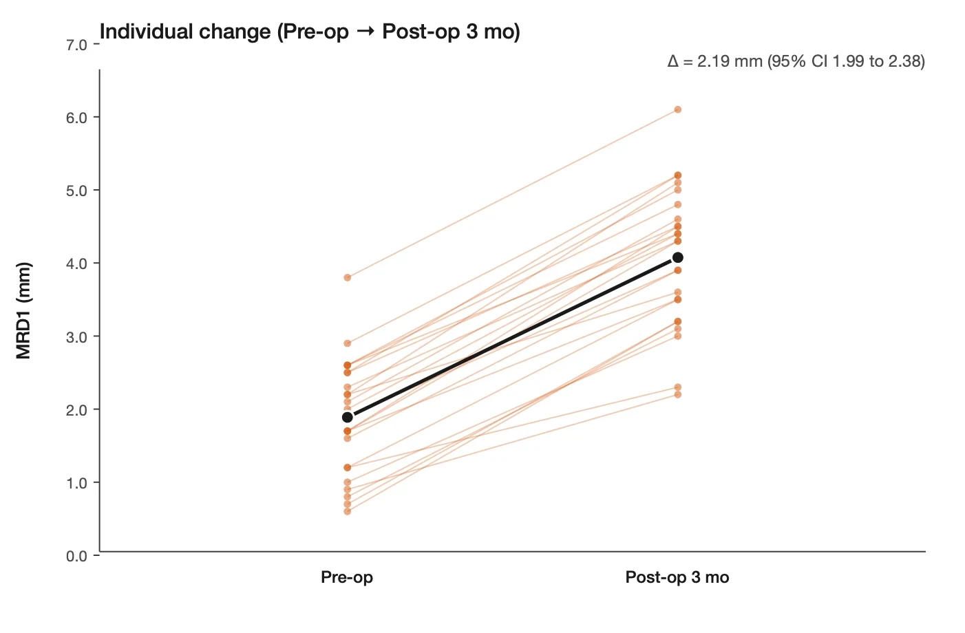
Before vs after
Did MRD1 change after ptosis repair?
Δ 2.19 mm 95% CI 1.99 to 2.38
Enlarge the Before vs after figure
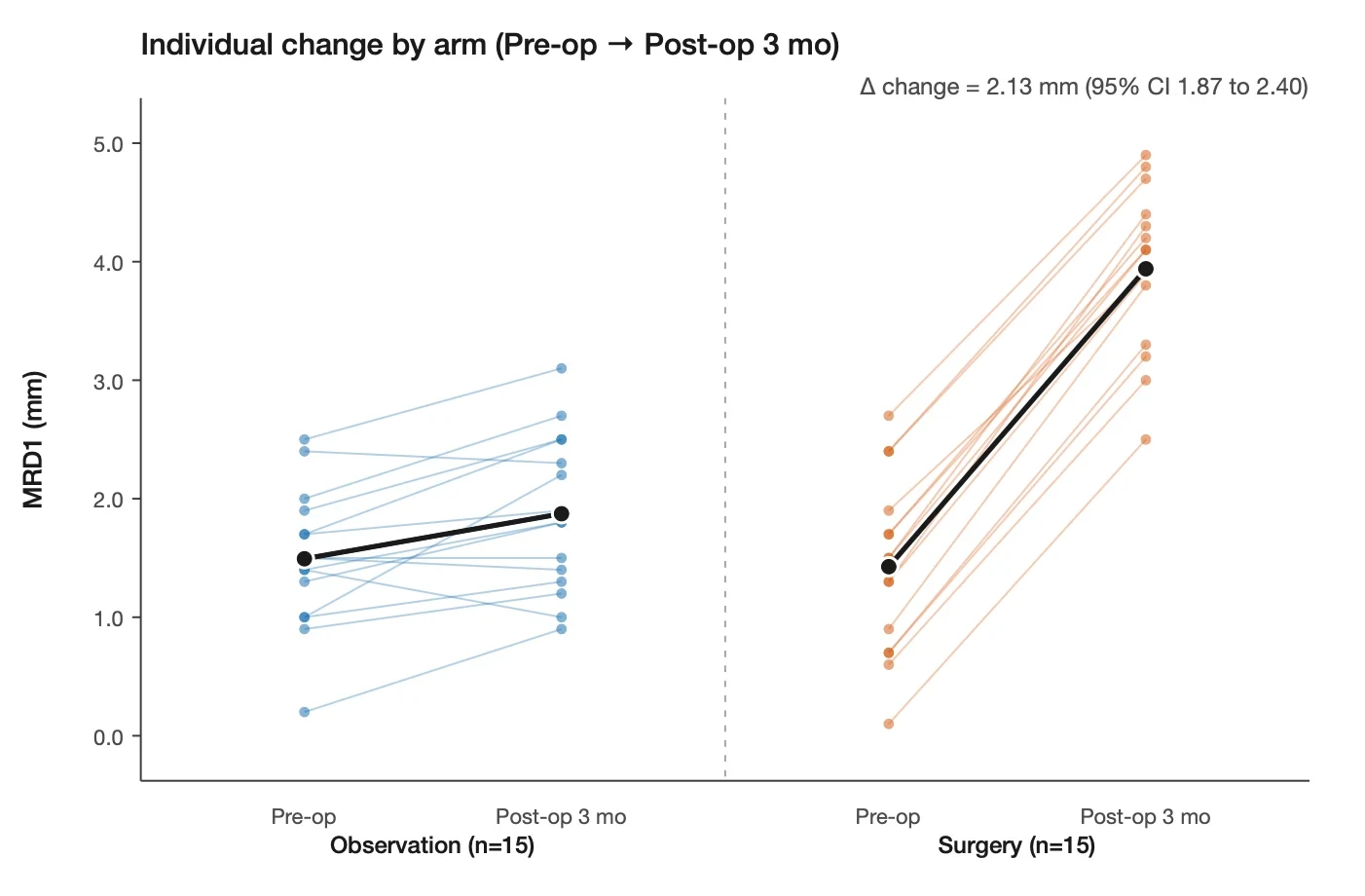
Before vs after, two groups
Did MRD1 change more after surgery than with observation?
Δ 2.13 mm 95% CI 1.87 to 2.40
Enlarge the Before vs after, two groups figure
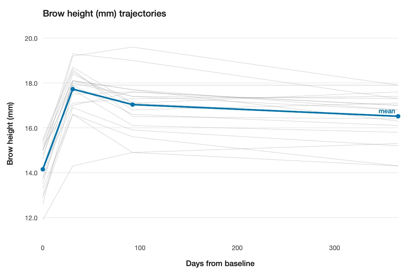
Longitudinal
How does brow height settle after a brow lift?
Δ 2.36 mm Month 12 · 95% CI 2.01 to 2.72
Enlarge the Longitudinal figure
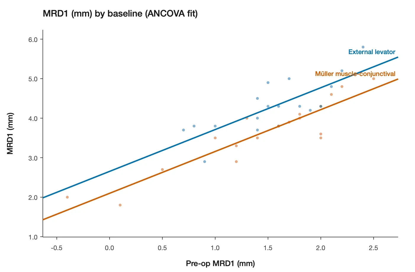
Two groups
Which lifts the lid more: levator advancement or Müller resection?
−0.55 mm 95% CI −0.82 to −0.29
Enlarge the Two groups figure
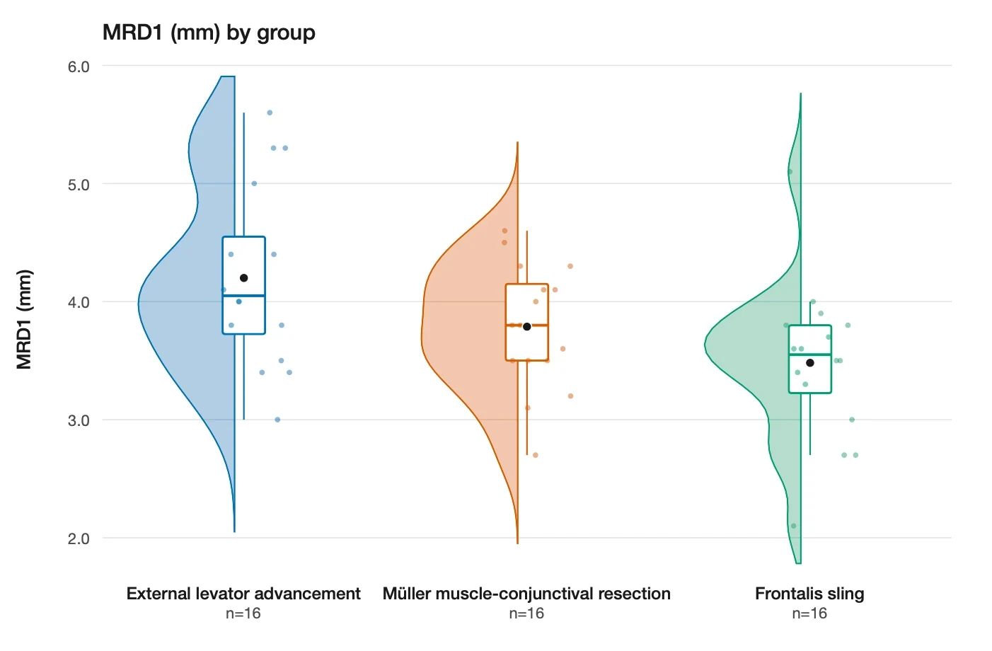
Three or more groups
Do three ptosis techniques give different results?
F 4.76 Global test · p = 0.01
Enlarge the Three or more groups figure
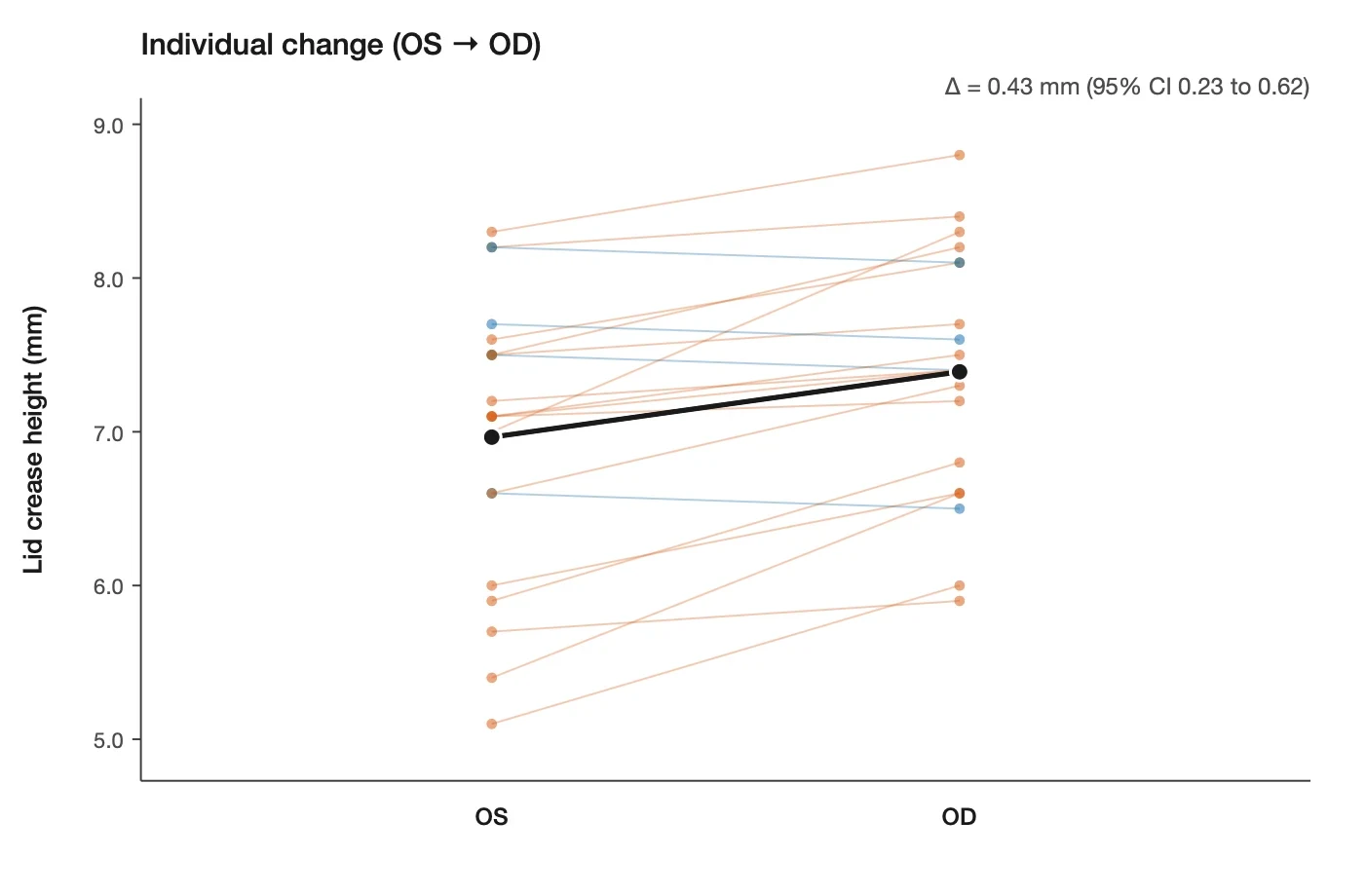
Fellow-eye
Does a new crease fixation differ from the standard one in the fellow eye?
0.43 mm 95% CI 0.23 to 0.62
Enlarge the Fellow-eye figure
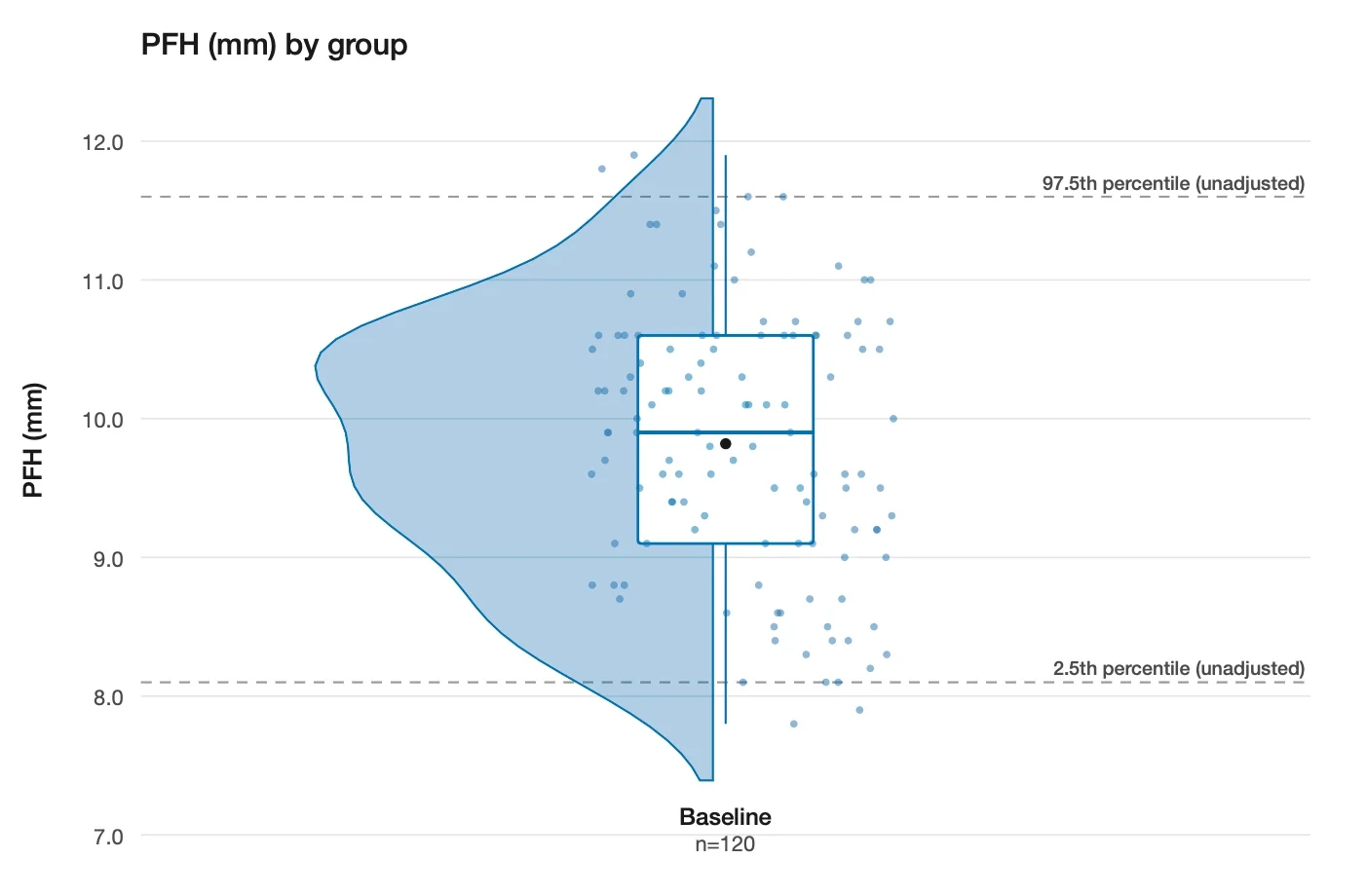
Normative
What is a normal palpebral fissure height?
PFH 9.82 mm Reference range 8.59 to 11.05
Enlarge the Normative figure
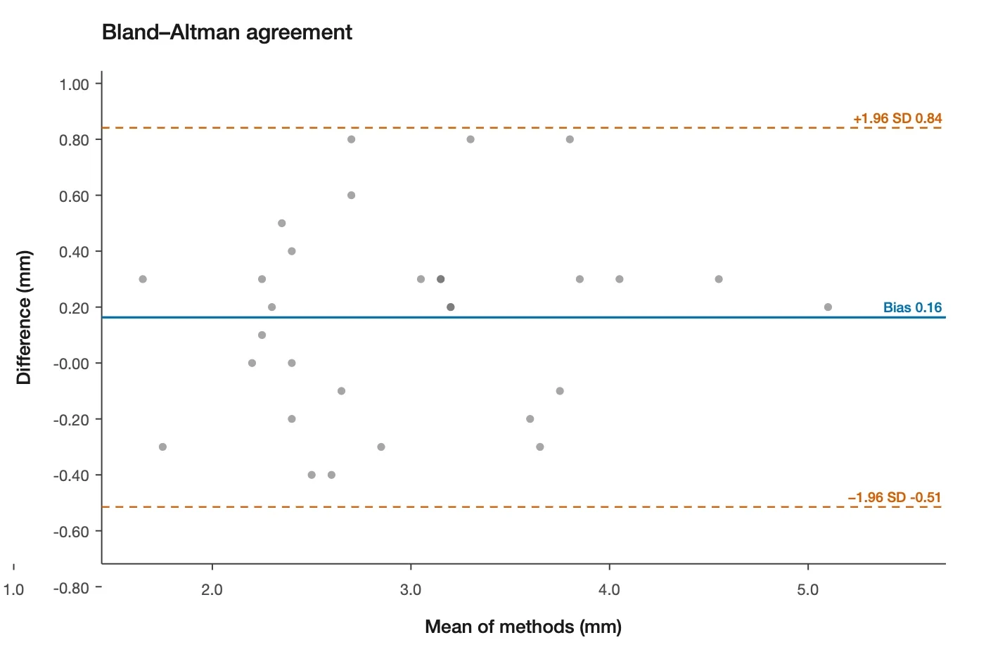
Reliability
Do manual and automated MRD1 measurements agree?
ICC 0.90 95% CI 0.77 to 0.95
Enlarge the Reliability figure
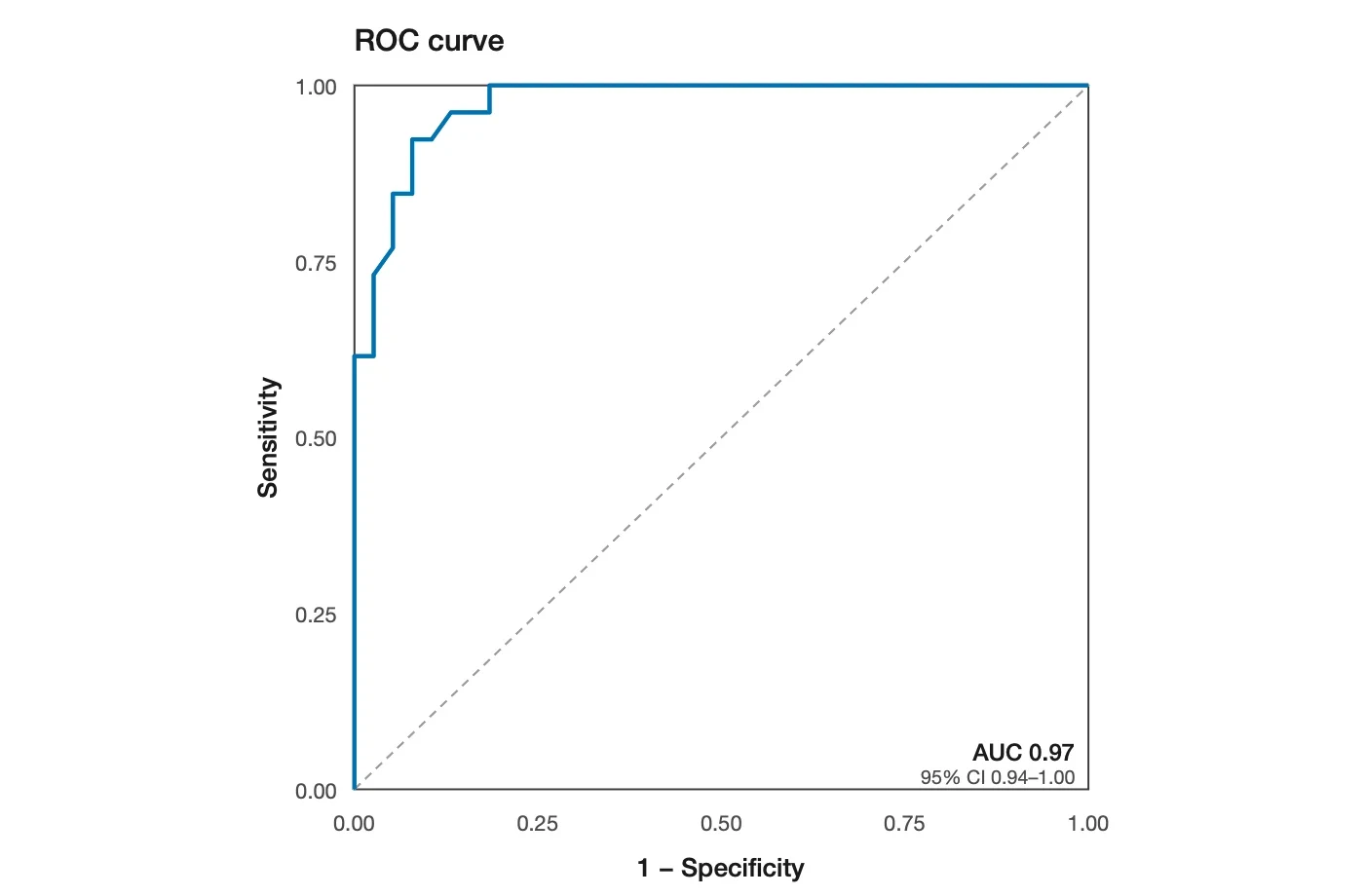
Diagnostic
How well does an AI score match surgeon grading of ptosis?
AUC 0.97 95% CI 0.94 to 1.00
Enlarge the Diagnostic figure
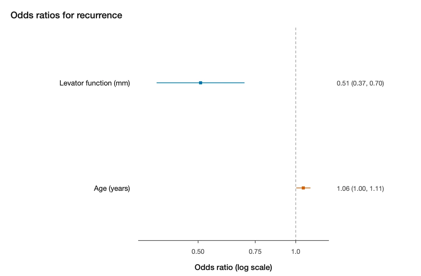
Prognostic
Does levator function predict success?
OR 0.51 Recurrence · 95% CI 0.37–0.70
Enlarge the Prognostic figurePick one of ten study designs, from before-and-after to diagnostic validation. The analysis follows from your question.
Every result is an effect estimate with a 95% confidence interval, not only a p-value.
The app flags small samples, unlocked endpoints, and data leakage, and says why.
Mixed models, ICC, Bland–Altman, ROC, and risk models are cross-validated against reference R implementations.
07 · Privacy
There is no OphthoRuler server. The developers never see your photographs, participants, or numbers.
AI models are run locally on your Mac or iPhone using Core ML. No image is uploaded to the internet to be analysed.
Photographs and study records are encrypted with AES-256-GCM using a password only you know.
All photos, AI models, and statistical analyses are carried out locally on your iPhone and Mac.
Mac and iPhone pair by QR code and sync over your own network, encrypted in transit. No cloud.
EXIF, GPS, and maker notes are removed on capture and import. A face is still identifiable, so this reduces risk but does not de-identify a photo.
You own your data. Your data and photos are never uploaded anywhere by OphthoRuler. All data is stored locally on your iPhone and Mac by default.
OphthoRuler is experimental research software. It has not been submitted for medical-device authorisation, is not certified against any privacy or health-data standard, and can be wrong.
06 · Questions
Researchers in oculoplastics, ophthalmology, and facial anthropometry who photograph eyes and need consistent, reproducible periocular measurements and a sound way to analyse them.
A Mac with macOS 15 or later for annotation, measurement, export, and analysis, and optionally an iPhone with iOS 18 or later for guided capture. The two sync directly over your local network.
The app reports every value with a propagated ± uncertainty and records how it was calibrated. Accuracy depends on calibration, head pose, lens and focus, and the landmarks themselves. It also depends on the quality of the segmentation, whether from the AI model you chose or from landmarks you edited by hand. Check the output, and edit landmarks where the model is wrong. As new and improved AI models are developed, they can be installed and used within OphthoRuler.
No. OphthoRuler is not a medical device and must not be used to diagnose, screen, monitor, or plan treatment. We built OphthoRuler to facilitate research studies in oculoplastic surgery. It is your responsibility to obtain REB/IRB approval to use OphthoRuler in your studies.
Only on your own devices, encrypted with your password. There is no cloud service and no developer access. Exports go only where you save them. This also means that if you lose your password we are unable to help you recover your data. Make sure you keep multiple backups in various formats and locations.
Yes. Measurements are built from landmarks in a visual editor (with a text view of the underlying formula) that you can inspect, duplicate, and edit, and measurement sets can be imported and exported. Bundled models ship with their licences and citations; adding and sharing your own models is coming during the beta.
OphthoRuler is in beta. Join the waitlist below and tell us a little about your research. We will be in touch as places open.
Beta · macOS 15+ and iOS 18+
Tell us a little about your research. We open places in small groups and will email you when yours is ready.